"International Journal of Applied Dental Sciences"
Vol-1, Issue-1
Three dimensional helical computed tomographic evaluation of two obturation techniques: An in vitro study
Author: Panna Mangat, Anil Dhingra, Sagarika Muni
Abstract:
Three Dimensional Helical Computed Tomographic Evaluation of Two Obturation Techniques: An In Vitro Study.
Aim: The aim of this study was to evaluate the adequacy of two obturation techniques, namely Calamus and Thermafil via volume rendering method utilizing a three dimensional helical computed tomography.
Materials and Method: Sixty freshly extracted single rooted teeth (maxillary first premolar) were collected and randomly allocated into two groups. Biomechanical preparation was done in all the teeth using rotary instruments. The teeth were placed in helical CT scanner and imaged before obturation. The teeth were then obturated utilizing following methods: Group 1- Calamus and group 2 - Thermafill. Evaluation of the volume of the pulp chamber and Gutta-percha after obturation was done via volume rendering technique, which reflects the adequacy of obturating system.
Results: There is a statistical difference between the Thermafill and Calamus with regards to the adequacy of obturation. The three dimensional obturating material Calamus have less volume inadequacy as compared to Thermafill.
Conclusion: The adequacy of obturation was better with Calamus as compared to Thermafill. But the research with regards to Calamus and Thermafill still continues in endodontics as three dimensional obturating material.

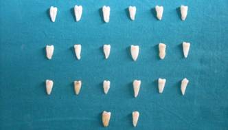
Fig: This figure represents specimens used in this study.
.Download Full Article: Click HereClick Hereee